Relating Cell Structure to Function through Coloring: Animal Cell Coloring Questions
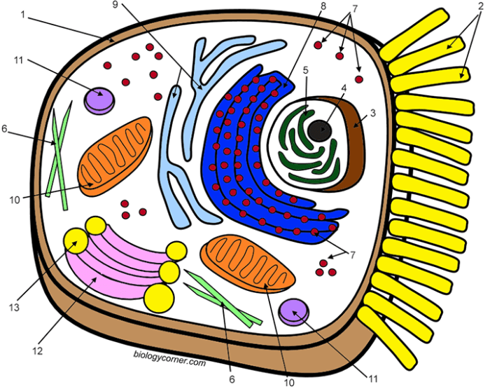
Animal cell coloring questions – Coloring activities offer a unique and engaging way to understand the intricate workings of an animal cell. By associating specific colors with different organelles, students can visually grasp the relationship between an organelle’s structure and its function within the complex cellular environment. This approach transforms abstract concepts into tangible representations, fostering a deeper understanding of cellular processes.
Shape and Structure Reflect Organelle Function
The shape and structure of each organelle are directly related to its specific role within the cell. For example, the highly folded inner membrane of the mitochondria, called cristae, significantly increases the surface area available for ATP production, the cell’s main energy source. The extensive network of the endoplasmic reticulum (ER), a continuous membrane system, facilitates protein synthesis and lipid metabolism.
Its folded structure maximizes its capacity for these crucial processes. Similarly, the spherical shape of the ribosomes optimizes their function in protein synthesis. Their compact structure allows for efficient binding to mRNA and tRNA molecules. The flattened, disc-like structure of the Golgi apparatus allows for the efficient processing, packaging, and transport of proteins and lipids. The large central vacuole in plant cells (though not in animal cells) provides structural support and stores water and nutrients, reflecting its function in maintaining cell turgor.
These examples highlight the principle that form follows function at the cellular level.
Tackling animal cell coloring questions can sometimes feel challenging, especially when identifying the various organelles. Fortunately, a helpful resource for clarifying these structures is readily available: you can find solutions to many common questions by checking out this useful worksheet with answers, animal cell coloring diagram worksheet answers. Reviewing these answers should help solidify your understanding and improve your ability to accurately answer future animal cell coloring questions.
Coloring Aids Visualization of Organelle Interconnectedness
A coloring activity can effectively illustrate the interconnectedness of organelles within a cell. For instance, coloring the endoplasmic reticulum (ER) one color and the Golgi apparatus another, but showing the connection between the two via a bridge of the same color, helps students visualize how proteins synthesized on the ER are transported to the Golgi for further processing. Similarly, demonstrating the close proximity of ribosomes to the ER visually reinforces their collaborative role in protein synthesis.
By using color-coding to show the flow of materials between organelles, the activity helps students understand the dynamic and integrated nature of cellular processes.
Addressing Potential Misconceptions about Animal Cell Structure
Coloring activities can effectively address common misconceptions about animal cell structure. For instance, students might initially perceive organelles as isolated entities rather than interconnected components of a dynamic system. The act of coloring the cell and its organelles, while highlighting their relationships, directly counters this misconception. Another common misconception involves the size and relative proportions of organelles. A carefully designed coloring sheet can accurately represent the relative sizes and spatial arrangements of different organelles, correcting any inaccurate preconceived notions.
Finally, the coloring activity can help clarify the distinction between different organelles, for example, the difference between the smooth and rough ER, based on the presence or absence of ribosomes.
Benefits of Using Color to Represent Cellular Processes, Animal cell coloring questions
Using color to represent different cellular processes within an animal cell enhances understanding and retention. For example, assigning a specific color to the process of protein synthesis (perhaps highlighting the ribosomes and ER in that color) makes it easier to follow this pathway visually. Similarly, different shades of a color could represent different stages of a process, such as the maturation of proteins as they move through the Golgi apparatus.
Color-coding helps students distinguish between different metabolic pathways and understand the complex interplay of cellular functions. The visual distinction created by color enhances memory and comprehension, facilitating learning.
A Day in the Life of an Animal Cell
It’s another busy day for Cellula, our typical animal cell! The mitochondria, our powerhouses (bright orange), are buzzing, generating ATP (represented by tiny yellow sparks) to fuel all our activities. The endoplasmic reticulum (a swirling blue network) is churning out proteins, while the ribosomes (tiny purple dots) diligently translate the genetic instructions. The Golgi apparatus (a stacked gold structure) efficiently packages and ships the proteins to their destinations. The lysosomes (small red spheres) diligently clean up cellular waste. The nucleus (a large purple sphere) safeguards our DNA, providing the blueprints for all our functions. The cell membrane (a vibrant green outer layer) regulates what enters and exits, ensuring we maintain a healthy internal environment. It’s a coordinated effort, a beautiful ballet of molecular machinery, all working together to keep Cellula thriving.
Beyond Basic Coloring
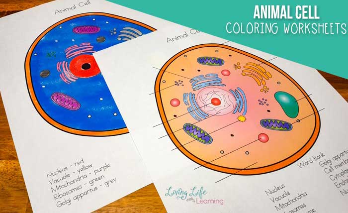
Taking your understanding of animal cells beyond simple coloring involves engaging in more interactive and analytical activities. These activities help solidify your knowledge of cell structures and their functions, moving from passive observation to active learning. The following activities offer a range of approaches to deepen your comprehension.
Labeling and Short Answer Questions
This advanced coloring activity enhances the basic coloring sheet by incorporating labels for each organelle and short answer questions that test comprehension of organelle function. For example, after coloring the Golgi apparatus, students could label it and then answer a question like: “Describe the role of the Golgi apparatus in protein modification and transport.” Similarly, labeling the mitochondria and answering a question about its role in cellular respiration would reinforce the connection between structure and function.
This method allows for immediate feedback and a more comprehensive understanding of the cell’s components.
Creating a 3D Animal Cell Model
A three-dimensional model provides a tactile and visually engaging way to learn about animal cell structure. Materials readily available at home or in a classroom can be used. For instance, a clear plastic bowl can represent the cell membrane. Different colored modeling clay or playdough can represent various organelles, such as the nucleus (a large sphere), the mitochondria (small oval shapes), the endoplasmic reticulum (a network of interconnected tubes), and the Golgi apparatus (flattened sacs).
Each organelle should be appropriately sized and positioned relative to others. This hands-on activity facilitates spatial understanding and reinforces the relative sizes and positions of organelles within the cell.
A Creative Coloring Project: Cell Mural or Diorama
A collaborative mural depicting an animal cell or an individual diorama showcasing a detailed representation of the cell can be highly engaging. The mural could feature large-scale drawings of organelles, each labeled and accompanied by a short description of its function. The diorama could use a variety of materials, such as cardboard boxes, construction paper, cotton balls (for the cytoplasm), and small plastic objects to represent organelles.
Students can work individually or in groups to create these projects, fostering teamwork and creativity while reinforcing their understanding of animal cell structure.
Integrating Coloring with Microscopes and Videos
Integrating coloring activities with other learning methods enhances learning outcomes. Observing prepared slides of animal cells under a microscope before coloring allows students to visually correlate the colored representation with the actual structures. Following this with educational videos explaining the function of each organelle further solidifies their understanding. This multi-sensory approach caters to different learning styles and provides a more holistic understanding of the subject.
Step-by-Step Guide for Detailed Animal Cell Illustration
Creating an accurate illustration requires a systematic approach.
- Sketching the Cell Membrane: Begin by sketching a roughly circular shape to represent the cell membrane. This is the outer boundary of the cell.
- Nucleus Placement: Position the nucleus, a large, centrally located, round or oval structure, within the cell. It should be relatively large compared to other organelles.
- Cytoplasm Representation: Lightly shade the area surrounding the organelles to represent the cytoplasm, the jelly-like substance filling the cell.
- Mitochondria Detailing: Draw numerous small, oval-shaped mitochondria scattered throughout the cytoplasm. These are the powerhouses of the cell.
- Endoplasmic Reticulum Network: Illustrate the endoplasmic reticulum as a network of interconnected, folded membranes. Distinguish between rough ER (studded with ribosomes) and smooth ER.
- Golgi Apparatus Depiction: Draw the Golgi apparatus as a stack of flattened sacs near the nucleus. This is involved in protein packaging and transport.
- Ribosomes and Vesicles: Add small dots (ribosomes) to the rough ER and depict small, membrane-bound vesicles throughout the cytoplasm. These transport materials within the cell.
- Lysosomes and Centrosomes: Include small, round lysosomes (involved in waste breakdown) and a pair of centrosomes near the nucleus (involved in cell division).
- Labeling and Color-Coding: Label each organelle clearly and use different colors to distinguish them visually. A key should accompany the illustration.
FAQ Corner
What are some common misconceptions about animal cell structure that coloring activities can help address?
Students often misrepresent the size and relative positions of organelles, or fail to grasp the interconnectedness of different cellular processes. Coloring activities can visually clarify these relationships.
How can I adapt animal cell coloring activities for different age groups?
Younger students can focus on basic shapes and colors, while older students can incorporate labeling, detailed drawings, and more complex projects.
What materials are needed for a basic animal cell coloring activity?
You will need printable worksheets, colored pencils or crayons, and potentially a reference image of an animal cell.
Are there online resources to help create animal cell coloring pages?
Yes, numerous websites and educational resources offer printable animal cell diagrams and coloring pages.

