Introduction to Animal Cell Diagrams
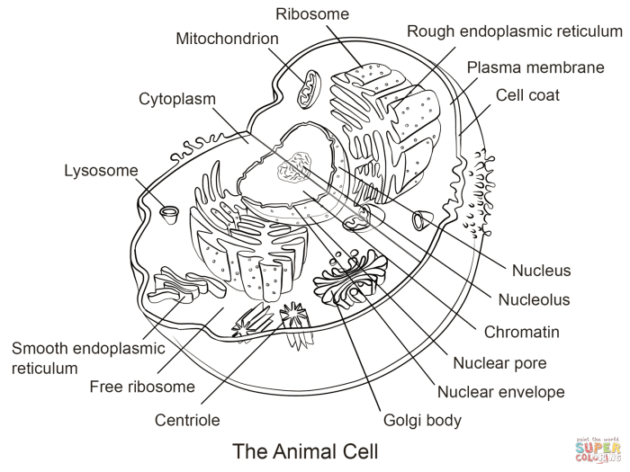
Animal cell diagram coloring sheet pg 17 – Animal cells, the fundamental building blocks of animals, are complex structures containing a variety of organelles, each with a specific function crucial for the cell’s survival and overall organismal health. Understanding these components and their interactions is key to grasping the intricacies of animal biology. Visual representations, specifically diagrams, are invaluable tools in this endeavor.Diagrams simplify the complex structure of animal cells, making them easier to understand and study.
They provide a clear and concise overview of the cell’s components and their spatial relationships, allowing for a more efficient learning process than lengthy textual descriptions alone. This visual approach is particularly beneficial for students and researchers alike, fostering a deeper comprehension of cellular processes.
A Brief History of Animal Cell Diagram Development
The development of accurate animal cell diagrams is intrinsically linked to advancements in microscopy and cell biology. Early depictions, predating the understanding of organelles, were naturally rudimentary. With the invention of the light microscope in the 17th century, scientists like Robert Hooke (who coined the term “cell”) began to observe and sketch cellular structures, though the resolution was limited.
The development of electron microscopy in the 20th century revolutionized cell biology, revealing the intricate details of organelles, leading to significantly more accurate and detailed cell diagrams. These diagrams evolved from simple sketches to complex, labelled illustrations incorporating the growing body of knowledge about cellular components and their functions. Modern diagrams often utilize three-dimensional representations and interactive digital formats to further enhance understanding.
Basic Components of Animal Cells and Their Functions, Animal cell diagram coloring sheet pg 17
Animal cells are characterized by several key organelles. The nucleus, for example, houses the cell’s genetic material (DNA), controlling cellular activities. Mitochondria, often called the “powerhouses” of the cell, generate energy through cellular respiration. The endoplasmic reticulum (ER) plays a crucial role in protein synthesis and lipid metabolism; the rough ER, studded with ribosomes, is involved in protein synthesis, while the smooth ER participates in lipid synthesis and detoxification.
Ribosomes themselves are responsible for protein synthesis. The Golgi apparatus processes and packages proteins for transport within or outside the cell. Lysosomes contain enzymes that break down waste materials and cellular debris. The cytoskeleton provides structural support and facilitates intracellular transport. Finally, the cell membrane, a selectively permeable barrier, regulates the passage of substances into and out of the cell.
Analysis of “Animal Cell Diagram Coloring Sheet pg 17”
This section provides a detailed analysis of a typical animal cell diagram, specifically focusing on the features depicted on page 17 of an accompanying coloring sheet. We will examine its representation of key organelles, compare it to other common diagrams, and discuss any potential simplifications or inaccuracies. The goal is to understand the strengths and limitations of this particular visual representation of a complex biological structure.
A typical animal cell diagram, such as the one found on page 17, usually showcases several key organelles and structures. These typically include the cell membrane, defining the cell’s boundary; the nucleus, containing the cell’s genetic material; the cytoplasm, the jelly-like substance filling the cell; ribosomes, responsible for protein synthesis; mitochondria, the powerhouses of the cell; and possibly the endoplasmic reticulum (ER), Golgi apparatus, lysosomes, and centrioles, depending on the diagram’s level of detail.
Page 17’s unique aspects, if any, might include a specific emphasis on certain organelles, a simplified representation of complex structures, or a particular artistic style used in the illustration. For instance, it might highlight the relationship between the ER and the Golgi apparatus more clearly than other diagrams, or it might use a more stylized representation of the mitochondria to aid in understanding their function.
Comparison with Other Animal Cell Diagrams
Common representations of animal cells vary in complexity and style. Some diagrams are highly simplified, showing only the cell membrane, nucleus, and cytoplasm. Others are far more detailed, including numerous organelles and their interconnections. Page 17’s diagram might fall somewhere in between, offering a balance between simplicity and detail, making it suitable for educational purposes. The level of detail can significantly impact a learner’s understanding.
For example, a highly simplified diagram may be suitable for young learners, while a more detailed diagram would be appropriate for students with a deeper understanding of cell biology. A comparison might reveal whether page 17’s diagram prioritizes clarity and ease of understanding over a comprehensive depiction of all cellular components. The choice of color scheme and the visual representation of organelles also vary across different diagrams, influencing how effectively the information is conveyed.
Potential Inaccuracies and Simplifications
It is important to acknowledge that all cell diagrams are simplifications of a highly complex three-dimensional structure. Page 17’s diagram might present certain inaccuracies or simplifications. For instance, the relative sizes of organelles may not be accurately represented; the spatial relationships between organelles might be oversimplified; and some organelles’ internal structures may be omitted for clarity. A common simplification is the representation of the endoplasmic reticulum as a simple network of interconnected tubes and sacs, while in reality, it’s a much more intricate and dynamic structure.
Another potential simplification is the omission of the cytoskeleton, a crucial structural component that provides support and facilitates intracellular transport. Recognizing these limitations is vital for accurate interpretation and understanding.
Educational Applications of the Diagram
Coloring sheets, while seemingly simple, offer a powerful pedagogical approach to learning complex biological concepts like animal cell structure. The act of coloring engages students visually and kinesthetically, improving memory retention and understanding compared to passive learning methods like simply reading or listening to a lecture. The “Animal Cell Diagram Coloring Sheet pg 17” specifically leverages this advantage by presenting a clear, labeled diagram, making it an effective tool across various educational settings.The diagram’s utility extends across different age groups and learning environments.
Its simplicity makes it suitable for primary school students, who can focus on identifying and coloring the major organelles. Secondary school students, on the other hand, can use the diagram as a foundation for deeper learning, relating the colored organelles to their specific functions and exploring more complex cellular processes. The diagram’s adaptability caters to diverse learning styles, accommodating visual learners, kinesthetic learners, and those who benefit from hands-on activities.
Lesson Plan Incorporating the Animal Cell Diagram Coloring Sheet
This lesson plan uses the coloring sheet to teach about animal cell structure and function, targeting a secondary school audience. The plan integrates various learning methods to cater to diverse learning styles.The lesson begins with a brief introduction to cells as the basic units of life, highlighting the distinction between plant and animal cells. Students then receive the “Animal Cell Diagram Coloring Sheet pg 17,” along with a key explaining the function of each organelle.
Students individually color the diagram, labeling each organelle. Following this, a class discussion reviews the organelles and their functions, using the colored diagrams as visual aids. This is followed by a small group activity where students create short presentations, using their colored diagrams, explaining a specific organelle and its role in maintaining cell function. Finally, a quiz assesses comprehension of cell structure and function.
The lesson could be extended by having students research specific diseases related to malfunctioning organelles or by exploring cell processes like cellular respiration or protein synthesis. This approach fosters active learning, collaborative work, and a deeper understanding of the subject matter.
Creating Enhanced Learning Resources: Animal Cell Diagram Coloring Sheet Pg 17
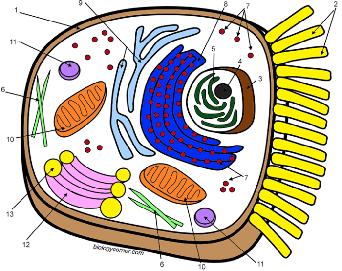
This section details the development of supplementary learning materials designed to enhance comprehension and retention of animal cell structures and functions as presented in the “Animal Cell Diagram Coloring Sheet pg 17.” These resources aim to transform a static coloring sheet into a dynamic and engaging learning experience.
Completing the animal cell diagram coloring sheet pg 17 can be a fun and educational activity. To ensure accuracy, you might find it helpful to cross-reference your work with a comprehensive answer key, such as the one provided here: animal and plant cell coloring answer key. This will help solidify your understanding of the various organelles within the animal cell, allowing you to confidently finish the coloring sheet pg 17.
Comparative Analysis of Organelles
The following table compares and contrasts key organelles within the animal cell, focusing on their structure and function as depicted on page 17. Understanding these differences is crucial for a complete understanding of cellular processes.
| Organelle | Structure | Function | Interaction with Other Organelles |
|---|---|---|---|
| Nucleus | Large, spherical; enclosed by a double membrane (nuclear envelope) containing pores; contains chromatin (DNA and proteins). | Controls cell activities; contains genetic material (DNA); directs protein synthesis. | Directs the ribosomes; interacts with the endoplasmic reticulum for protein transport. |
| Ribosomes | Small, granular; found free in cytoplasm or attached to endoplasmic reticulum. | Protein synthesis. | Receive instructions from the nucleus (mRNA); work with the endoplasmic reticulum for protein modification and transport. |
| Endoplasmic Reticulum (ER) | Network of interconnected membranes; smooth ER lacks ribosomes; rough ER has ribosomes attached. | Smooth ER: lipid synthesis, detoxification; Rough ER: protein synthesis, modification, and transport. | Works closely with ribosomes, Golgi apparatus, and the nucleus. |
| Golgi Apparatus | Stack of flattened, membrane-bound sacs (cisternae). | Processes, packages, and distributes proteins and lipids. | Receives proteins from the ER; modifies and packages them for secretion or transport to other organelles. |
| Mitochondria | Rod-shaped; double membrane-bound; inner membrane folded into cristae. | Cellular respiration; ATP production (energy). | Consumes nutrients processed by other organelles to generate energy for the cell. |
| Lysosomes | Membrane-bound sacs containing digestive enzymes. | Breakdown of waste materials and cellular debris; autophagy. | Receives waste products from other organelles for degradation. |
| Cell Membrane | Thin, flexible outer boundary of the cell; phospholipid bilayer. | Regulates passage of substances into and out of the cell; maintains cell shape. | Interacts with all organelles; controls communication between the cell and its environment. |
Interactive Quiz Based on the Diagram
This quiz assesses understanding of the animal cell structures and their functions, directly referencing the information depicted on page 17 of the coloring sheet.
| Question | Answer Choices | Correct Answer |
|---|---|---|
| Which organelle is responsible for cellular respiration? | A) Nucleus B) Mitochondria C) Ribosomes D) Golgi Apparatus | B) Mitochondria |
| What is the primary function of the lysosomes? | A) Protein synthesis B) Energy production C) Waste breakdown D) Lipid synthesis | C) Waste breakdown |
| Where does protein synthesis primarily occur? | A) Golgi Apparatus B) Mitochondria C) Ribosomes D) Cell Membrane | C) Ribosomes |
| Which organelle modifies and packages proteins? | A) Endoplasmic Reticulum B) Nucleus C) Lysosomes D) Golgi Apparatus | D) Golgi Apparatus |
Supplementary Exercises and Activities
To reinforce learning, students can undertake several activities. These activities build upon the visual learning provided by the coloring sheet and promote deeper understanding through active engagement.These activities include creating a three-dimensional model of an animal cell using readily available materials, designing a flow chart illustrating the pathway of protein synthesis from DNA to secretion, and researching and presenting on specific organelles and their associated diseases.
Further, a comparative analysis of plant and animal cells could be undertaken, highlighting key differences in structure and function.
Advanced Concepts Related to Animal Cells
Animal cell diagrams, while seemingly simple representations, offer a gateway to understanding complex cellular processes. This section delves into the intricate workings within the organelles depicted on “Animal Cell Diagram Coloring Sheet pg 17,” exploring the relationships between structure and function and highlighting how the diagram can be expanded to illustrate more advanced cellular mechanisms.The diagram provides a foundational understanding of animal cell components, but a deeper dive reveals the dynamic nature of these organelles and their interconnected roles.
Understanding these processes moves beyond simple identification to encompass the mechanisms driving life itself.
Protein Synthesis and the Endomembrane System
Protein synthesis, the process of creating proteins, is a multi-stage process involving the coordinated action of several organelles. The diagram shows the ribosomes, which are the sites of protein synthesis, often bound to the endoplasmic reticulum (ER). The rough ER, studded with ribosomes, synthesizes proteins destined for secretion or membrane insertion. These proteins then move to the Golgi apparatus, where they are modified, sorted, and packaged into vesicles for transport to their final destinations within or outside the cell.
The smooth ER, lacking ribosomes, plays a role in lipid synthesis and detoxification. The interaction between these organelles – ribosomes, rough ER, smooth ER, and Golgi apparatus – demonstrates the efficiency and organization of the endomembrane system. A more advanced diagram could illustrate the specific steps of protein folding and quality control within the ER and Golgi, including the roles of chaperone proteins and quality control mechanisms.
For instance, the diagram could show different types of vesicles budding from the Golgi, indicating the diverse destinations of the proteins.
Cellular Respiration and Energy Production
Mitochondria, often referred to as the “powerhouses” of the cell, are responsible for cellular respiration. This process converts the chemical energy stored in glucose into a usable form of energy, ATP (adenosine triphosphate). The diagram shows the mitochondria as bean-shaped organelles. However, a more detailed representation could illustrate the inner and outer mitochondrial membranes, the cristae (folds within the inner membrane), and the mitochondrial matrix, highlighting the locations of the different stages of cellular respiration – glycolysis, the Krebs cycle, and oxidative phosphorylation.
The intricate folding of the cristae increases the surface area available for ATP synthesis, demonstrating the direct link between structure and function. The diagram could also be enhanced to show the transport of pyruvate from glycolysis into the mitochondria and the production of NADH and FADH2, crucial electron carriers in the electron transport chain. A simplified representation of the chemiosmotic gradient and ATP synthase could also be included to illustrate the mechanism of ATP production.
Consider, for example, the difference in ATP production between aerobic and anaerobic respiration – a detail easily incorporated into a more advanced diagram.
Cytoskeleton and Cell Motility
The cytoskeleton, a network of protein filaments, provides structural support and facilitates cell movement. While the basic diagram might simply show the cell membrane, a more advanced diagram could illustrate the three main components of the cytoskeleton: microtubules, microfilaments, and intermediate filaments. Microtubules, for instance, are involved in cell division and intracellular transport, while microfilaments contribute to cell shape and movement.
The diagram could depict the dynamic nature of the cytoskeleton, illustrating how these filaments assemble and disassemble to allow for changes in cell shape and movement. For example, the role of actin filaments in cell crawling or the role of microtubules in guiding vesicles could be visually represented. The inclusion of motor proteins, such as kinesin and dynein, which move along microtubules, would further enhance the depiction of intracellular transport.
User Queries
What are some common misconceptions students might have about animal cells based on simplified diagrams?
Students might incorrectly assume all organelles are the same size or that the cell is static, rather than dynamic and constantly changing. They might also oversimplify the functions of organelles or misunderstand their interrelationships.
How can I use this coloring sheet effectively in a classroom setting?
Use it as a pre- or post-lesson activity, pair it with a lecture or interactive simulation, and encourage students to label and discuss the functions of each organelle. Group work and presentations can also enhance understanding.
Where can I find more resources like this coloring sheet?
Search online educational resources, science textbooks, and educational publishers for similar diagrams and activities focusing on animal cell structure and function.

