Key Organelles: Animal Cell Plant Cell Coloring Sheet
Animal cell plant cell coloring sheet – Cells, the fundamental units of life, exhibit remarkable diversity in structure and function, particularly between plant and animal cells. Understanding the roles of key organelles is crucial to grasping the differences in their capabilities and overall biological processes. This section will delve into a comparison and contrast of these essential cellular components.
Cell Wall and Cell Membrane: A Functional Comparison
The cell wall and cell membrane are both crucial for maintaining cell integrity, but they differ significantly in their structure and function. The cell wall, a rigid outer layer found only in plant cells, provides structural support and protection. It is primarily composed of cellulose, a complex carbohydrate that forms a strong, yet permeable, barrier. The cell membrane, present in both plant and animal cells, is a selectively permeable phospholipid bilayer that regulates the passage of substances into and out of the cell.
While the cell wall provides structural rigidity, the cell membrane controls the cell’s internal environment through selective permeability. The cell membrane is dynamic, allowing for processes like endocytosis and exocytosis, while the cell wall provides a more static, protective barrier.
Chloroplast Function in Plant Cells
Chloroplasts are unique to plant cells and are the sites of photosynthesis, the process by which light energy is converted into chemical energy in the form of glucose. These organelles contain chlorophyll, a green pigment that absorbs light energy, and a complex system of internal membranes where the light-dependent and light-independent reactions of photosynthesis occur. The glucose produced during photosynthesis serves as the primary source of energy for the plant and is used to build other organic molecules.
Without chloroplasts, plants would be unable to produce their own food and would rely entirely on external sources of energy.
Vacuole Function in Plant and Animal Cells
Vacuoles are membrane-bound organelles found in both plant and animal cells, but their size and function differ significantly. In animal cells, vacuoles are generally small and numerous, playing roles in intracellular transport, storage of waste products, and maintaining cell turgor pressure to a lesser extent. Plant cells, however, possess a large, central vacuole that occupies a significant portion of the cell’s volume.
This central vacuole plays a crucial role in maintaining turgor pressure, providing structural support, storing water, nutrients, and waste products, and regulating the cell’s internal environment. The size difference reflects the differing needs of plant and animal cells; plants rely heavily on the central vacuole for structural support and water storage, whereas animal cells have other mechanisms for these functions.
Nucleus Structure and Function
The nucleus is a prominent organelle found in both plant and animal cells, serving as the control center of the cell. It is enclosed by a double membrane called the nuclear envelope, which contains pores that regulate the passage of molecules between the nucleus and the cytoplasm. Inside the nucleus, the genetic material, DNA, is organized into chromosomes.
The nucleus is responsible for controlling gene expression, DNA replication, and cell division. While the basic structure and function of the nucleus are similar in both plant and animal cells, the size and organization of the chromosomes may vary slightly.
Organelle Comparison: Plant vs. Animal Cells
The following table summarizes the functions of various organelles in plant and animal cells, highlighting their similarities and differences.
| Organelle | Animal Cell Function | Plant Cell Function | Differences |
|---|---|---|---|
| Cell Membrane | Regulates transport of substances; maintains cell integrity | Regulates transport of substances; maintains cell integrity | Similar function, but plant cells also have a cell wall |
| Cell Wall | Absent | Provides structural support and protection | Present only in plant cells |
| Chloroplasts | Absent | Site of photosynthesis | Present only in plant cells |
| Vacuole | Small, involved in transport and waste storage | Large central vacuole, maintains turgor pressure, stores water and nutrients | Significant size and functional differences |
| Mitochondria | Site of cellular respiration (ATP production) | Site of cellular respiration (ATP production) | Similar function in both cell types |
| Nucleus | Controls gene expression, DNA replication, and cell division | Controls gene expression, DNA replication, and cell division | Similar function, but may vary slightly in size and chromosome organization |
| Ribosomes | Protein synthesis | Protein synthesis | Similar structure and function |
| Endoplasmic Reticulum | Protein and lipid synthesis, transport | Protein and lipid synthesis, transport | Similar function, but may differ in extent of development |
| Golgi Apparatus | Protein modification and packaging | Protein modification and packaging | Similar function |
Coloring Sheet Design and Educational Value
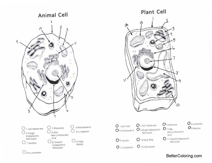
Coloring sheets offer a fun and engaging way to learn about complex biological concepts like cell structure. By visually representing the components of animal and plant cells, these sheets can aid comprehension and retention, particularly for younger learners. The act of coloring itself can improve fine motor skills and hand-eye coordination, adding a multi-faceted benefit to the learning experience.
Animal Cell Coloring Sheet Design
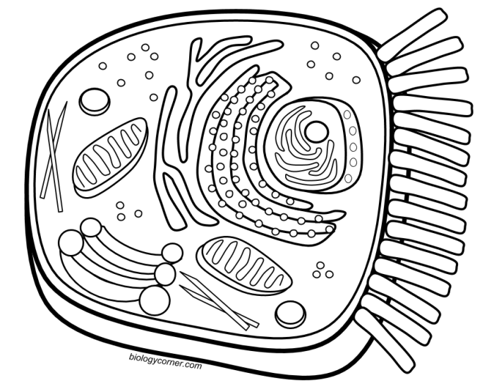
The animal cell coloring sheet should depict a typical animal cell, perhaps a generalized mammalian cell. The cell membrane should be shown as a thin, continuous outer boundary, colored perhaps a light blue or beige. The nucleus, a large, centrally located organelle, can be a darker shade, like purple or dark pink. The nucleolus, within the nucleus, could be a smaller, contrasting circle.
The rough endoplasmic reticulum (RER) can be depicted as a network of interconnected membranes studded with ribosomes (small dots), colored in a light green. The smooth endoplasmic reticulum (SER), lacking ribosomes, could be a lighter, more pastel green. Mitochondria, the “powerhouses” of the cell, can be illustrated as bean-shaped structures in a reddish-brown or orange. The Golgi apparatus can be depicted as a stack of flattened sacs, perhaps a light yellow or tan.
Lysosomes, small spherical organelles involved in waste breakdown, can be colored a dark blue or purple. Finally, the cytoplasm, the jelly-like substance filling the cell, can be a pale yellow or a light beige. Each organelle should be clearly labeled with its name.
Plant Cell Coloring Sheet Design
The plant cell coloring sheet should highlight the key differences from an animal cell. The cell wall, a rigid outer layer, should be a darker green or brown, clearly distinct from the cell membrane (light green). The large central vacuole, a fluid-filled sac that occupies much of the cell’s volume, can be a light purple or blue. Chloroplasts, responsible for photosynthesis, should be depicted as oval-shaped green organelles, possibly with internal structures (grana) suggested by lighter green shading.
The other organelles (nucleus, mitochondria, endoplasmic reticulum, Golgi apparatus) can be colored similarly to the animal cell sheet, maintaining visual consistency. Again, all organelles should be clearly labeled.
Engaging Coloring Sheets for Different Age Groups
Adapting the coloring sheets for different age groups is crucial for maximizing engagement. For younger children (preschool – early elementary), larger, simpler drawings with fewer organelles are ideal. Bright, bold colors and possibly even simple illustrations of the cell’s functions (like a tiny sun for chloroplasts) can be incorporated. Older children (upper elementary – middle school) can handle more detailed diagrams with a larger number of labeled organelles.
Interactive elements, such as requiring them to research and add further details or create their own simple diagrams based on the coloring sheet, can increase engagement.
Educational Benefits of Cell Structure Coloring Sheets
Coloring sheets provide a hands-on, visual learning experience that reinforces abstract concepts. The process of identifying, labeling, and coloring each organelle strengthens memory and understanding of cell structure and function. It also fosters a deeper appreciation for the complexity of even the simplest life forms. The visual nature of the activity makes it accessible to a wide range of learning styles, catering to both visual and kinesthetic learners.
Integrating Coloring Sheets into a Lesson Plan
Introduction
Use the coloring sheet as a pre-activity to introduce the topic of cell structure. Students can color the sheet before a formal lecture or discussion.
Reinforcement
After a lesson on cells, the coloring sheet can serve as a review activity to solidify understanding and identify areas needing further clarification.
Assessment
The completed coloring sheets can be used as a simple formative assessment to gauge student comprehension of cell organelles and their functions.
Creative Extension
Students can create their own diagrams, labeling additional organelles or drawing comparisons between animal and plant cells.
Differentiation
Provide varying levels of complexity for different students, perhaps simplifying the drawings or providing fill-in-the-blank labels for younger students.
Advanced Cell Structures and Processes (Optional)
This section delves into more complex aspects of cell biology, providing a deeper understanding of the intricate workings within both plant and animal cells. We will explore the structural support provided by the cytoskeleton, the energy-generating processes of photosynthesis and cellular respiration, and the mechanisms of cell division. Finally, we will examine specialized cells and their unique adaptations.
The Cytoskeleton’s Role in Maintaining Cell Shape and Function, Animal cell plant cell coloring sheet
The cytoskeleton is a dynamic network of protein filaments extending throughout the cytoplasm. It acts as an internal scaffolding, providing structural support and maintaining cell shape. This network is crucial for various cellular processes, including cell division, intracellular transport, and cell motility. Three main types of filaments comprise the cytoskeleton: microtubules (long, hollow tubes), microfilaments (thin, solid rods), and intermediate filaments (fibrous proteins).
Microtubules are involved in chromosome segregation during cell division and intracellular transport, while microfilaments contribute to cell shape changes and movement. Intermediate filaments provide mechanical strength and anchor organelles. The cytoskeleton’s ability to constantly rearrange itself allows cells to adapt to changing conditions and perform their diverse functions.
Animal and plant cell coloring sheets are a fantastic way to learn cell structure. Understanding the differences between the two is crucial, and a helpful resource for accurately completing your animal cell sheet is the answer key available at animal cell coloring biology corner answer key. This will allow you to compare your work and reinforce your understanding before moving on to more complex aspects of plant cell coloring.
Ultimately, both sheets offer a valuable visual learning experience.
Photosynthesis in Plant Cells
Photosynthesis is the process by which plants convert light energy into chemical energy in the form of glucose. This occurs within chloroplasts, organelles containing chlorophyll, the green pigment that absorbs light energy. The process can be summarized in two main stages: the light-dependent reactions and the light-independent reactions (Calvin cycle). In the light-dependent reactions, light energy is used to split water molecules, releasing oxygen and producing ATP and NADPH, energy-carrying molecules.
The light-independent reactions utilize the ATP and NADPH to convert carbon dioxide into glucose. The overall equation for photosynthesis is: 6CO 2 + 6H 2O + Light Energy → C 6H 12O 6 + 6O 2.
Cellular Respiration in Animal and Plant Cells
Cellular respiration is the process by which cells break down glucose to release energy in the form of ATP. This occurs in the mitochondria, often referred to as the “powerhouses” of the cell. Both plant and animal cells utilize cellular respiration, although plants also produce glucose through photosynthesis. The process involves several stages: glycolysis (occurs in the cytoplasm), the Krebs cycle (occurs in the mitochondrial matrix), and oxidative phosphorylation (occurs in the inner mitochondrial membrane).
Oxidative phosphorylation generates the majority of ATP through a process called chemiosmosis, where the movement of protons across the inner mitochondrial membrane drives ATP synthesis. The overall equation for cellular respiration is: C 6H 12O 6 + 6O 2 → 6CO 2 + 6H 2O + ATP.
Mitosis and Meiosis: A Comparison
Mitosis and meiosis are two types of cell division. Mitosis is a process of cell duplication, or proliferation, resulting in two identical daughter cells. Meiosis, on the other hand, is a type of cell division that results in four daughter cells, each with half the number of chromosomes as the parent cell. This reduction in chromosome number is essential for sexual reproduction.
Both processes involve similar stages (prophase, metaphase, anaphase, telophase), but meiosis includes two rounds of division (meiosis I and meiosis II), resulting in the reduction of chromosome number. Plant cells form a cell plate during cytokinesis (the final stage of cell division), whereas animal cells form a cleavage furrow.
Specialized Animal and Plant Cells and Their Unique Adaptations
The following table showcases examples of specialized cells and their unique adaptations:
| Cell Type | Organism | Function | Unique Adaptation |
|---|---|---|---|
| Neuron | Human | Transmission of nerve impulses | Long axon for rapid signal transmission |
| Red Blood Cell | Human | Oxygen transport | Biconcave shape and lack of nucleus for efficient oxygen carrying |
| Guard Cell | Plant | Regulation of stomata opening and closing | Ability to change shape to control gas exchange |
| Root Hair Cell | Plant | Water and nutrient absorption | Long, thin extensions to increase surface area for absorption |
Visual Representations and Extensions
Creating effective visual representations and extending the coloring sheet activity into larger projects significantly enhances learning and engagement. High-quality visuals aid comprehension, while broader projects encourage deeper exploration of cell biology. The following sections detail how to achieve this.
Alt Text Descriptions for Cell Images
Providing accurate alt text for images is crucial for accessibility and . Below are detailed descriptions suitable for use with images of animal and plant cells.
Animal Cell Alt Text: A microscopic image of an animal cell, showing a round or irregular shape. Key organelles are visible, including a centrally located nucleus (the cell’s control center), numerous smaller ribosomes (responsible for protein synthesis), a complex network of endoplasmic reticulum (involved in protein and lipid metabolism), a Golgi apparatus (modifying and packaging proteins), and mitochondria (the powerhouses of the cell).
The cytoplasm, a jelly-like substance filling the cell, surrounds these organelles. The cell membrane, a thin outer boundary, encloses the entire structure.
Plant Cell Alt Text: A microscopic image of a plant cell, typically rectangular or polygonal in shape. The cell is enclosed by a rigid cell wall, providing structural support. Inside the wall, a cell membrane encloses the cytoplasm, which contains organelles similar to those in an animal cell, including a nucleus, ribosomes, endoplasmic reticulum, Golgi apparatus, and mitochondria. However, plant cells also feature prominent chloroplasts (responsible for photosynthesis), a large central vacuole (maintaining turgor pressure and storing water and nutrients), and plasmodesmata (small channels connecting adjacent plant cells).
Infographic Design for Comparing Animal and Plant Cells
A visually appealing infographic effectively compares and contrasts animal and plant cells. The following steps Artikel the creation of such an infographic.
Creating an effective infographic involves a structured approach to visual communication. Careful consideration of layout, color schemes, and clear labeling are essential for conveying complex information concisely.
- Layout: Divide the infographic into two main sections, one for animal cells and one for plant cells. Use a clear and consistent layout, perhaps a side-by-side comparison or a top-bottom arrangement.
- Visual Elements: Use simple, clear illustrations of both cell types. Employ color-coding to highlight key organelles and their functions. Consider using different shapes and sizes to represent the relative sizes of organelles.
- Text: Keep the text concise and easy to read. Use bullet points or short sentences to describe the key features of each organelle. Include a brief summary of the differences between animal and plant cells.
- Color Scheme: Choose a color scheme that is visually appealing and easy on the eyes. Use contrasting colors to highlight important information. Maintain consistency throughout the infographic.
- Data Representation: Use charts or graphs to represent quantitative differences between animal and plant cells, such as the size of the vacuole or the number of chloroplasts.
Extending the Coloring Sheet Activity
The coloring sheet can be the foundation for more extensive projects that reinforce learning.
Extending the basic coloring sheet activity allows students to engage with the material more deeply and demonstrate their understanding in creative ways.
- Presentation: Students can create presentations summarizing their understanding of animal and plant cells, using the colored sheet as a visual aid. The presentation can include additional information about cell functions and processes.
- 3D Model: Students can construct 3D models of animal and plant cells using various materials like clay, foam, or even recycled items. This allows for a hands-on, tactile learning experience.
- Comparative Essay: Students can write a comparative essay detailing the similarities and differences between animal and plant cells, drawing on the information gained from the coloring sheet and additional research.
Essential FAQs
Are these coloring sheets suitable for all age groups?
Yes, with minor adjustments. Simpler versions can be created for younger children, while more detailed sheets can challenge older students.
Where can I find printable versions of these coloring sheets?
Printable versions can be easily generated from digital files created using common graphics software. Many educational websites also offer free printable cell coloring sheets.
What other activities can complement the use of these coloring sheets?
Building 3D models of cells, creating presentations, or researching specific organelles would be excellent supplementary activities.

