Coloring Activities
Animal cell coloring pdf – Alright, class, let’s get this party started! We’ve got the intro and outro all set, so it’s time to dive headfirst into the awesome world of animal cell coloring. Think of this worksheet as your very own VIP backstage pass to the inner workings of a cell – it’s gonna be legen…wait for it…dary!This coloring worksheet is designed to be both fun and educational, making learning about animal cell structure as easy as pie.
We’re talking about transforming a potentially dry topic into a vibrant, engaging experience. It’s the ultimate cell-ebration!
Animal Cell Coloring Worksheet Design
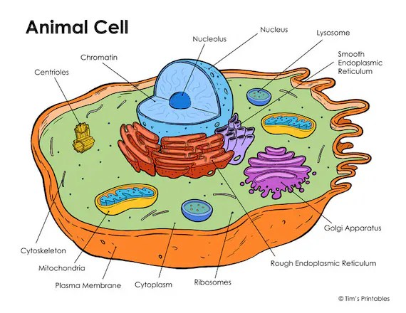
This worksheet will feature a large, central circle representing the cell membrane. Imagine it as a bouncy castle, keeping all the cell’s important parts safely inside. Within this circle, we’ll strategically place the major organelles, each clearly labeled for easy identification. Think of it as a cell’s “who’s who” – each organelle gets its moment in the spotlight.
We’ll have a nucleus, looking like a giant control center, a Golgi apparatus resembling a stack of pancakes, mitochondria shaped like little beans, and the endoplasmic reticulum, a network of tubes and sacs, snaking its way through the cell. Ribosomes, lysosomes, and the cytoplasm will fill in the spaces, creating a bustling, vibrant city within the cell membrane.
The overall design will be clear, uncluttered, and visually appealing, like a mini-masterpiece waiting to be colored.
Color Key and Organelle Assignments
To make things super clear, we’ll provide a handy color key. This key will assign a specific color to each organelle, ensuring consistency and aiding in understanding. For example, the nucleus could be a bold purple, representing its role as the cell’s brain. The mitochondria, the powerhouses, could be a vibrant red, reflecting their energy production. The Golgi apparatus could be a sunny yellow, and the endoplasmic reticulum a cool blue.
This color-coding system helps kids (and adults!) visually differentiate between organelles and better remember their functions. Think of it as a cell-based color-by-numbers, but way cooler.
Educational Value of Coloring Worksheets
Coloring isn’t just for kids; it’s a powerful learning tool that taps into multiple learning styles. This worksheet provides a hands-on, visual approach to understanding animal cell structure, making learning more engaging and memorable. The act of coloring reinforces the names and locations of organelles, improving retention. The color-coding adds an extra layer of visual organization, making it easier to distinguish and recall each organelle’s function.
Understanding animal cell structure can be enhanced through the use of an animal cell coloring pdf. This activity provides a visual and engaging way to learn. For a related activity that might be helpful before diving into the intricacies of cell biology, consider exploring simpler illustrations; you might find suitable options at animal coloring sheets to print to build foundational skills.
Returning to the animal cell coloring pdf, remember that accurate coloring helps solidify understanding of organelle placement and function.
It’s like a cheat code for memorization, but instead of unlocking extra lives, you unlock cellular knowledge! This method is particularly effective for visual learners, turning a potentially abstract concept into a tangible, colorful experience. It’s a win-win – learning and fun combined! It’s the ultimate educational power-up!
Advanced Cell Representations
Level up your cell diagram game! Forget basic coloring – we’re talking about creating seriously awesome, realistic representations of animal cells. Think less crayon scribbles and more high-def 3D renderings. We’re diving into techniques that’ll make your cell pop off the page (or screen!).This section explores methods to showcase the three-dimensional structure of an animal cell, moving beyond simple coloring to create truly impressive visuals.
We’ll cover shading and texture to add realism, discuss accurately representing organelle size and placement, and finally, show you how to construct a compelling cutaway diagram.
Three-Dimensional Cell Structure Representation
Creating a three-dimensional representation of an animal cell goes beyond flat coloring. Imagine you’re a microscopic filmmaker, capturing the cell’s intricate architecture. One effective method is to use overlapping circles and ovals of varying sizes and shades to suggest depth. For example, the nucleus could be represented as a large, slightly shaded sphere, with the nucleolus as a smaller, darker sphere nestled within.
Ribosomes, depicted as tiny dots, could be clustered around the endoplasmic reticulum, visualized as a network of interconnected tubes and sacs, varying in shades to show the difference between rough and smooth ER. The Golgi apparatus could be illustrated as stacked, flattened sacs, with the edges slightly darker to create a sense of depth. By layering these shapes and employing varying shades and textures, you can build a visually compelling three-dimensional representation.
Shading and Texture for Realism, Animal cell coloring pdf
Adding shading and texture is like applying makeup to your cell – it brings it to life! Think about the way light hits a real-world object. Use lighter shades to represent areas where light would directly hit, and darker shades for areas in shadow. For example, the concave surface of a mitochondrion can be highlighted with a lighter shade, while the shadowed inner folds receive darker tones.
Textures can be implied through the use of stippling (dots) or hatching (lines) to suggest the rough surface of the endoplasmic reticulum or the granular appearance of the cytoplasm. The use of gradients can also enhance the three-dimensionality of organelles. Consider the smooth, almost glassy appearance of the cell membrane, contrasting with the more textured appearance of the nucleus.
These subtle details greatly enhance the realism of your diagram.
Relative Sizes and Positions of Organelles
Accuracy is key! It’s not just about drawing organelles; it’s about getting their relative sizes and positions right. The nucleus, typically the largest organelle, should dominate the center. Mitochondria should be numerous and scattered throughout the cytoplasm. The endoplasmic reticulum forms an extensive network, extending throughout the cell. Visualizing the spatial relationships between organelles is crucial for understanding cellular processes.
Think of it like designing a miniature city; each organelle has its specific location and function. A well-executed diagram accurately reflects this spatial organization, making it easier to understand the complex interactions within the cell. For instance, you could show the proximity of ribosomes to the rough ER, highlighting their role in protein synthesis.
Creating a Cutaway Diagram
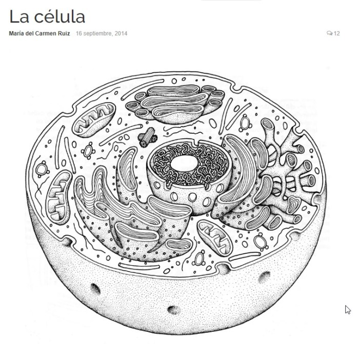
A cutaway diagram is like slicing open the cell to reveal its inner workings. It’s a fantastic way to showcase the internal structures and their relationships in a clear and engaging manner. Start by drawing the outer cell membrane as a complete oval. Then, strategically remove a portion of the cell membrane to expose the internal organelles. This allows you to clearly display the intricate arrangement of the organelles without the clutter of overlapping structures.
Different organelles can be highlighted using varying colors and textures, further emphasizing their unique characteristics. Imagine a cross-section of an apple; you can see the different layers and components clearly. A cutaway diagram of an animal cell works similarly, allowing for a clearer understanding of its internal structure and the spatial relationships between organelles. You could, for example, show a section through the nucleus, revealing the nucleolus and chromatin.
Creating a PDF for Download: Animal Cell Coloring Pdf
Yo, future cell biologists! Let’s get this animal cell coloring PDF ready for prime time. We’re talking slick, downloadable goodness that’ll have kids (and adults!) coloring away like it’s the latest TikTok trend. This section breaks down how to make that happen.Creating a printable PDF from your awesome animal cell diagram is easier than you think. It’s all about getting that digital masterpiece into a format everyone can access and print without losing any of its vibrant detail.
We’ll cover the steps, software, and tips to make sure your PDF is totally rad.
Software Options for PDF Creation and Editing
Several software options can help you create a killer PDF. Adobe Acrobat Pro is the industry standard, offering tons of features for professional-looking results. Think of it as the Beyoncé of PDF software – powerful, polished, and a little pricey. However, if you’re on a budget, free options like LibreOffice Draw or Google Docs can get the job done, offering surprisingly robust features for creating and editing PDFs.
For simpler tasks, even your operating system’s built-in print-to-PDF function might suffice. Choosing the right software depends on your budget and the complexity of your design.
Steps to Create a Printable PDF
First, you’ll need your digital animal cell diagram. Make sure it’s high-resolution; we’re aiming for crisp, clear lines and vibrant colors. Then, open your chosen software. Import your image, and arrange it on the page. If you’re using a program like Adobe Acrobat, you can add text elements like a title, labels for the cell organelles, and a legend.
Finally, export your document as a PDF. Make sure to choose a high-quality setting to preserve image resolution. Think of this step as carefully curating your masterpiece – every detail counts!
Ensuring Image Quality and Clarity
Image quality is king! A blurry, pixelated cell diagram is a major buzzkill. To avoid this, start with a high-resolution image. At least 300 DPI (dots per inch) is recommended for print. When saving your PDF, select a high-quality setting – typically “Print” or a similar option that prioritizes image fidelity over file size. Think of it like this: would you rather watch a grainy, low-res movie or a crystal-clear 4K masterpiece?
The choice is clear.
Organizing the PDF Layout
Think of your PDF like a perfectly curated album. You want a killer title page to grab attention. Then, the main event: the coloring page itself, featuring your detailed animal cell diagram. Finally, include a key or legend that clearly identifies each organelle. This helps users understand what they’re coloring.
A well-organized PDF is user-friendly and enhances the overall experience. This is your chance to show off your design skills and create a truly engaging learning tool. It’s like planning the perfect Instagram feed – visually appealing and informative!

